Independent and reproducible hippocampal radiomic biomarkers for multisite Alzheimer’s disease: diagnosis, longitudinal progress and biological basis
Kun Zhao1,2,3, Yanhui Ding3, Ying Han4,17,18,19, Yong Fan5, Aaron F. Alexander-Bloch6, Tong Han7, Dan Jin1,8, Bing Liu1,8,9, Jie Lu10, Chengyuan Song11, Pan Wang12,13, Dawei Wang14, Qing Wang14, Kaibin Xu1, Hongwei Yang10, Hongxiang Yao15, Yuanjie Zheng3, Chunshui Yu16, Bo Zhou13, Xinqing Zhang4, Yuying Zhou12, Tianzi Jiang1,8,9, Xi Zhang13*, Yong Liu1,8,9*
1Brainnetome Center & National Laboratory of Pattern Recognition, Institute of Automation, Chinese Academy of Sciences, Beijing, China;
2School of Biological Science and Medical Engineering, Beihang University, Beijing, China;
3School of Information Science and Engineering, Shandong Normal University, Ji’nan, China;
4Department of Neurology, Xuanwu Hospital of Capital Medical University, Beijing, China;
5Department of Radiology, Perelman School of Medicine, University of Pennsylvania, Philadelphia, USA;
6Department of Psychiatry, Yale University School of Medicine, New Haven, CT, USA;
7Department of Radiology, Tianjin Huanhu Hospital, Tianjin, China;
8School of Artificial Intelligence, University of Chinese Academy of Sciences, Beijing, China;
9Center for Excellence in Brain Science and Intelligence Technology, Institute of Automation, Chinese Academy of Sciences, Beijing, China;
10Department of Radiology, Xuanwu Hospital of Capital Medical University, Beijing, China;
11Department of Neurology, Qilu Hospital of Shandong University, Ji’nan, China;
12Department of Neurology, Tianjin Huanhu Hospital, Tianjin, China;
13Department of Neurology, The Secondary Medical Center, National Clinical Research Center for Geriatric Disease, Chinese PLA General Hospital, Beijing, China;
14Department of Radiology, Qilu Hospital of Shandong University, Ji’nan, China;
15Department of Radiology, The Secondary Medical Center, National Clinical Research Center for Geriatric Disease, Chinese PLA General Hospital, Beijing, China;
16Department of Radiology, Tianjin Medical University General Hospital, Tianjin, China;
17Center of Alzheimer’s Disease, Beijing Institute for Brain Disorders, Beijing, China;
18Beijing Institute of Geriatrics, Beijing, China;19National Clinical Research Center for Geriatric Disorders, Beijing, China;
Abstract
Hippocampal morphological change is one of the main hallmarks of Alzheimer's disease (AD). However, whether hippocampal radiomic features are robust as predictors of progression from mild cognitive impairment (MCI) to AD dementia and whether these features provide any neurobiological foundation remains unclear. The primary aim of this study was to verify whether hippocampal radiomic features can serve as robust MRI markers for AD. Multivariate classifier-based SVM analysis provided individual-level predictions for distinguishing AD patients (n=261) from normal controls (NCs; n=231) with an accuracy of 88.21% and intersite cross-validation. Further analyses of a large, independent ADNI dataset (n=1228) reinforced these findings. In MCI groups, a systemic analysis demonstrated that the identified features were significantly associated with clinical features (e.g., apolipoprotein E (APOE) genotype, polygenic risk scores, cerebrospinal fluid (CSF) Aβ, CSF Tau), and longitudinal changes in cognition ability; more importantly, the radiomic features had a consistently altered pattern with changes in the MMSE scores over 5 years of follow-up. These comprehensive results suggest that hippocampal radiomic features can serve as robust biomarkers for clinical application in AD/MCI, and further provide evidence for predicting whether an MCI subject would convert to AD based on the radiomics of the hippocampus. The results of this study are expected to have a substantial impact on the early diagnosis of AD/MCI.
Key words: Hippocampal radiomic features; Multisite Alzheimer’s disease MRI; Independent cross-validation; Brain biomarker; Biological basis
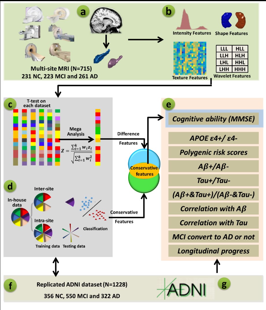
 | Figure 1. Schematic of the data analysis pipeline. (a) Automatic segmentation of the hippocampus in the individual space with the LLL method. (b) Computation of radiomic features. (c) T-test between NC and AD and mega-analysis to identify differences at the multicenter level. (d) Classification analysis between AD and NC using intersite and intrasite crossvalidation to test the generalizability of the results. (e) Correlation between MMSE and radiomic features (and the decision value of the subject to the classification plane). (f) Validation of the ADNI dataset for using ADNI as training data with in-house data as testing data as well as using in-house as training data with ADNI data as testing data to test the generalizability of the results. (g) Relationship between radiomic features and clinical information (e.g., cognitive ability, APOE genetics, Aβ, Tau, and longitudinal changes in the cognitive abilities in MCI). |
Article Link: https://www.sciencedirect.com/science/article/pii/S2095927320302140
