A dedicated eight-channel receive RF coil array for monkey brain MRI at 9.4T
Mingyan Li1 | Yu Li1 | Jin Jin 1,2 | Zhengyi Yang 1,3 | Baogui Zhang 3 | Yanyan Liu 3 | Ming Song 3,4,5 | Craig Freakly 1 | Ewald Weber 1 | Feng Liu 1 | Tianzi Jiang 3,4,5,6,7,8 | Stuart Crozier 1
1School of Information Technology and Electrical Engineering, The University of Queensland, Brisbane, Queensland, Australia
2Siemens Healthcare Pty. Ltd., Bowen Hills QLD, 4006, Australia
3Brainnetome Center, Institute of Automation, Chinese Academy of Sciences, Beijing, China
4National Laboratory of Pattern Recognition, Institute of Automation, Chinese Academy of Sciences, Beijing, China
5University of Chinese Academy of Sciences, Beijing, China
6CAS Center for Excellence in Brain Science and Intelligence Technology, Institute of Automation, Chinese Academy of Sciences, Beijing, China
7Key Laboratory for NeuroInformation of Ministry of Education, School of Life Science and Technology, University of Electronic Science and Technology of China, Chengdu, China
8Queensland Brain Institute, University of Queensland, Brisbane, Queensland, Australia
Abstract
The neuroimaging of nonhuman primates (NHPs) realised with magnetic resonance imaging (MRI) plays an important role in understanding brain structures and functions, as well as neurodegenerative diseases and pathological disorders. Theoretically, an ultrahigh field MRI (≥7 T) is capable of providing a higher signal-to-noise ratio (SNR) for better resolution; however, the lack of appropriate radiofrequency (RF) coils for 9.4 T monkey MRI undermines the benefits provided by a higher field strength. In particular, the standard volume birdcage coil at 9.4 T generates typical destructive interferences in the periphery of the brain, which reduces the SNR in the neuroscience-focused cortex region. Also, the standard birdcage coil is not capable of performing parallel imaging. Consequently, extended scan durations may cause unnecessary damage due to overlong anaesthesia. In this work, assisted by numerical simulations, an eight-channel receive RF coil array was specially designed and manufactured for imaging NHPs at 9.4 T. The structure and geometry of the proposed receive array was optimised with numerical simulations, so that the SNR enhancement region was particularly focused on monkey brain. Validated with rhesus monkey and cynomolgus monkey brain images acquired from a 9.4 T MRI scanner, the proposed receive array outperformed standard birdcage coil with higher SNR, mean diffusivity and fractional anisotropy values, as well as providing better capability for parallel imaging.
Keywords: applications, array coils, computational, electromagnetics, diffusion-weighted imaging, MR engineering, neurological, parallel imaging
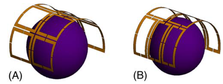
 | Figure. (A) 2 × 4 semi-open loop coil array. (B) 3–2-3 semiopen loop coil array |
